Skip to the main content ↓
★★★★★ 100% recommended 17 Facebook reviews

Surgery isn't your only option.

Fibroids, an enlarged prostate, varicose veins, thyroid nodules and tumours — treated through a 1–2 mm incision, under local anaesthetic. Most patients go home the same day.

2014
Practising since
5,000+
Procedures performed
100%
Recommended
HOW IT WORKS

Three steps, through a 1–2 mm incision.

Live imaging guides a catheter through your own blood vessels to the exact source of the problem — under local anaesthetic.

01 · NAVIGATE

Reaching the source

A 1–2 mm incision, not open surgery. A guidewire finer than a pencil lead follows your own blood vessels to the exact spot.

02 · TREAT

Targeted embolization

Microspheres released at the catheter tip block the blood supply feeding the problem — starved of flow, it shrinks.

03 · RESTORE

Flow restored

Where a vessel is narrowed, a stent opens it exactly where it is needed — and relief typically follows.

Every treatment is guided by live imaging, under local anaesthesia — most patients go home the same day.

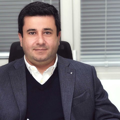
Dr. Agathangelou Marios, interventional radiologist
THE DOCTOR

Board-certified interventional radiologist. Practising since 2014.

Dr. Agathangelou Marios practises in Nicosia and Limassol, specialising in minimally invasive, image-guided treatments performed through a 1–2 mm incision — from vascular interventions to complex tumour ablations.

Board certified
Nicosia & Limassol
24 h emergency line
WHAT INTERVENTIONAL RADIOLOGY IS READ THE FULL PROFILE
WHAT THIS MEANS FOR YOU

Compared with open surgery.

IMAGE-GUIDED
OPEN SURGERY
Entry
1–2 mm incision
Surgical incision
Anaesthesia
Local ± sedation
Usually general
Hospital stay
Same day / 1 night
Several days
Recovery
Days
Weeks
Scar
None
Surgical scar

Indicative, general comparison — suitability of each technique is always assessed case-by-case.

WHY NO SURGERY
IN PRACTICE

What precision looks like.

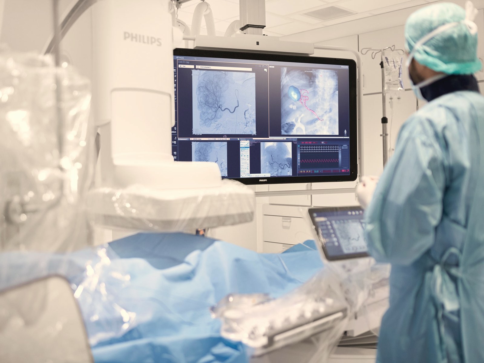
Image-guided procedure at the clinic
IMAGING GUIDED
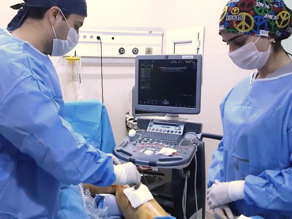
Varicose-vein procedure at the clinic
EVLA · VARICOSE VEINS
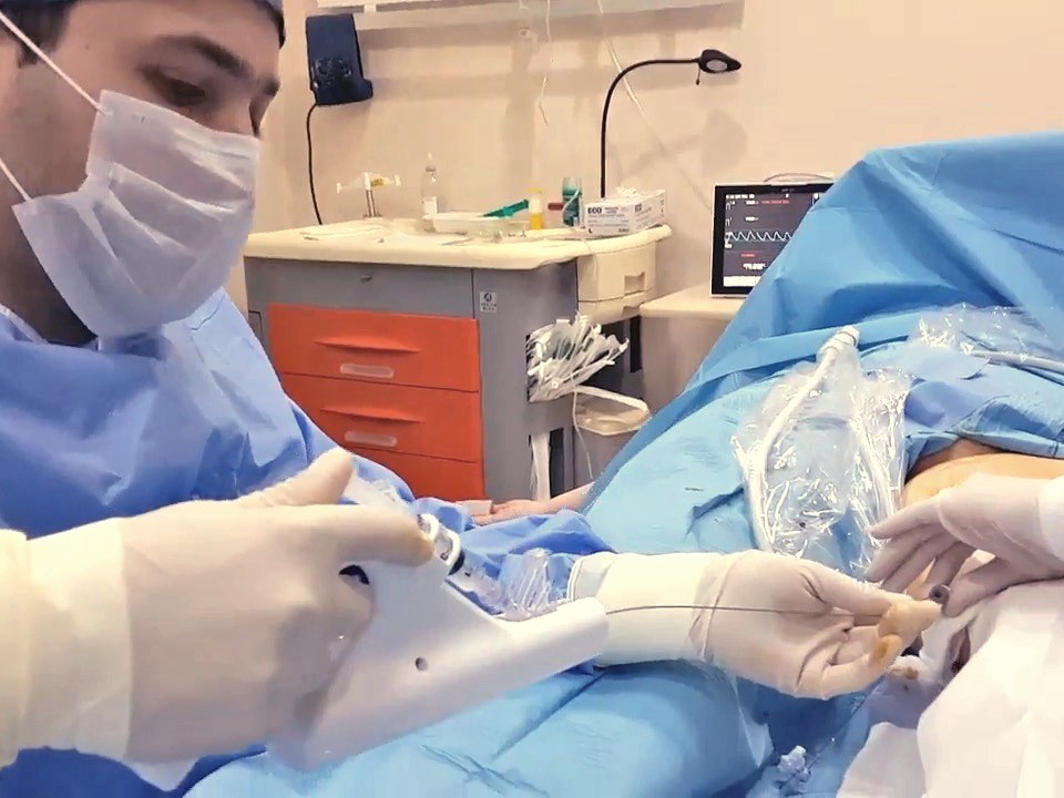
The 1–2 mm access point of an image-guided procedure
1–2 mm ACCESS
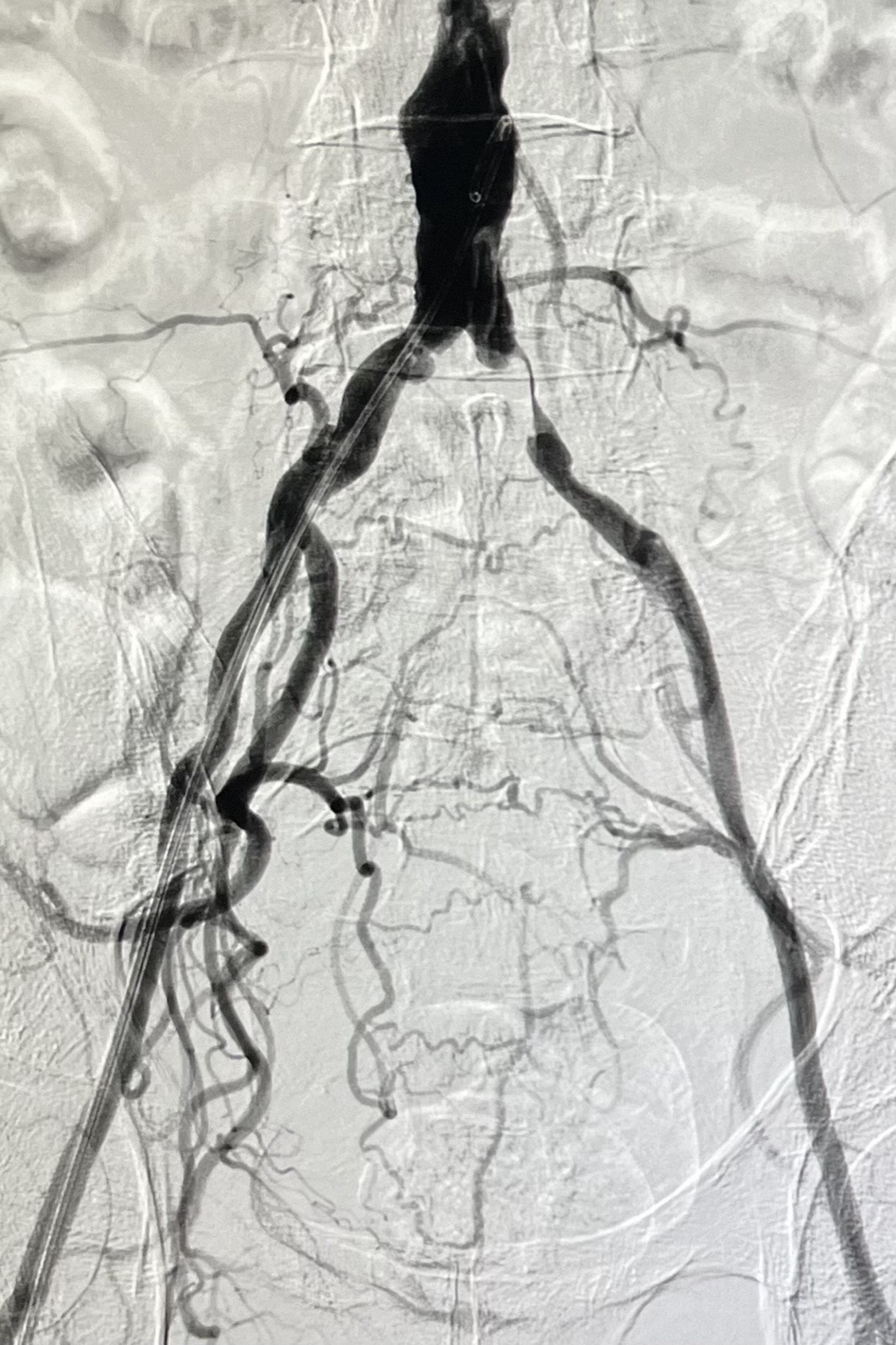
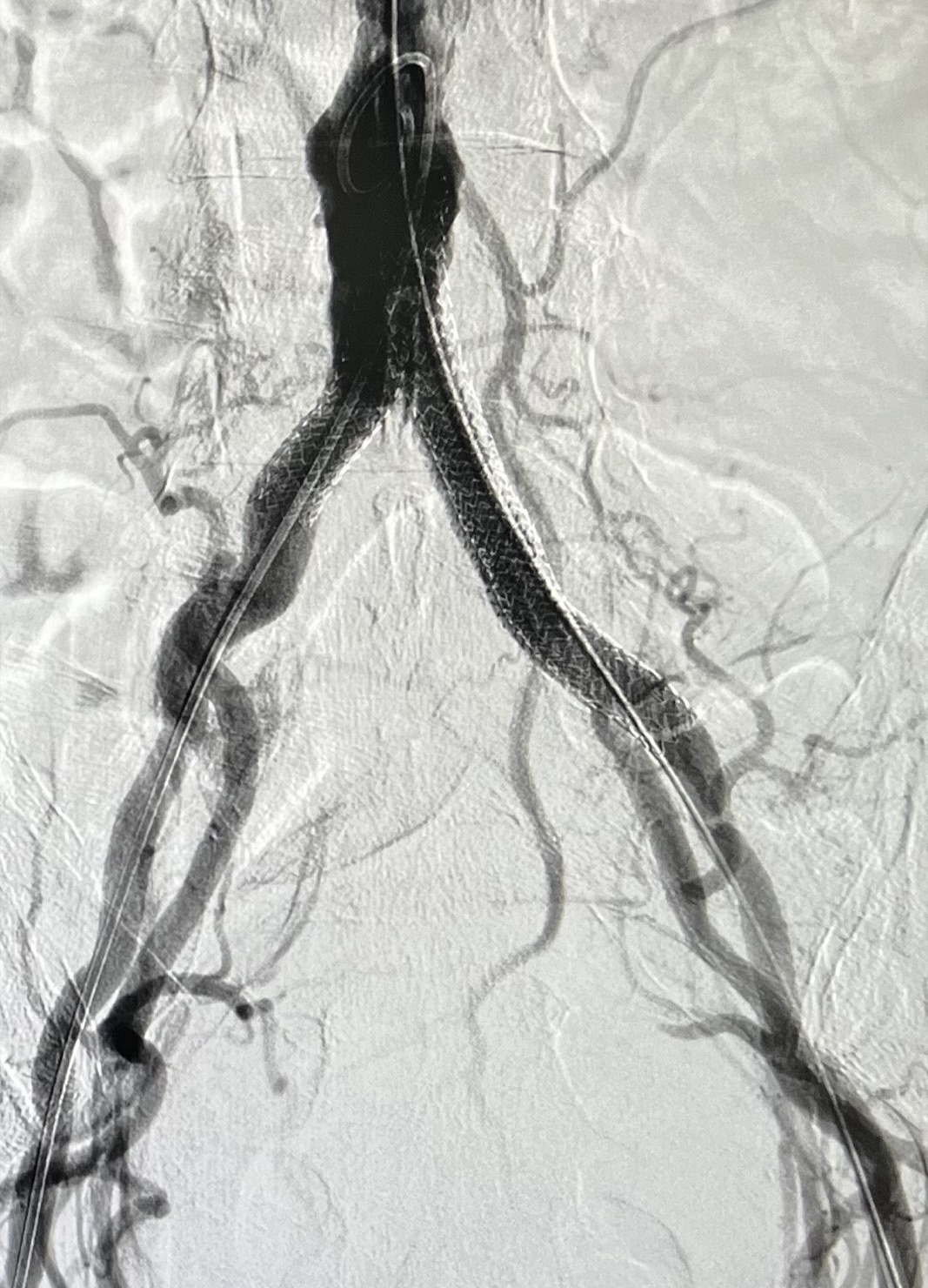
Digital subtraction angiogram of aorto-iliac stenosis
DSA · AORTO-ILIAC STENOSIS (BEFORE)
Digital subtraction angiogram after aorto-iliac stent placement
DSA · AORTO-ILIAC STENOSIS (AFTER)

DSA IMAGING SHOWN FOR PATIENT EDUCATION · INDIVIDUAL RESULTS VARY

“The procedure was quick, surprisingly painless, and my recovery was remarkably smooth. I was back to my normal routine within a week.”
MARIA K. · UTERINE FIBROID EMBOLIZATION · NICOSIA
“Dr Agathangelou took the time to thoroughly explain every step of the procedure and answered all my questions with patience.”
JOHN P. · PROSTATE ARTERY EMBOLIZATION · LIMASSOL

PUBLISHED WITH WRITTEN PATIENT CONSENT · INDIVIDUAL RESULTS VARY

READ ALL PATIENT REVIEWS
NEXT STEP

Not sure if you are a candidate?

Send your case for an opinion, or call the clinic directly — and if this isn’t right for you, we’ll say so. Consultations in Nicosia and Limassol.